
Moorthy A 1*, Jibi Paul 1, G. Muthuraj 1
Authors:
1 Faculty of Physiotherapy, DR MGR Educational and Research Institute, Velappanchavadi, Chennai, India
Corresponding Author:
1* Faculty of Physiotherapy, DR MGR Educational and Research Institute, Velappanchavadi, Chennai, India. Email: moorthympt@yahoo.co.in
ABSTRACT
Background of the study: Knee joint stiffness is one of the commonest complications in patients who had fractures in the femur and upper tibia. Stiffness, restrict range of motion of joint caused by soft tissue tightness and intra articular adhesions. The aim of study is to understand the effects of Muscle Energy Techniques on early knee joint mobilization to improve the range of motion by reducing post traumatic stiffness.
Methodology: A pretest-post test control group design was used for this study. Thirty patients from orthopedic physical therapy outpatient department of Sri Gokulam hospital, Salem were selected for this study and equally divided into two groups. Patients in group-A (Experimental) received wax therapy, static quadriceps exercise, active assisted mobilization and muscle energy techniques. Patients in group-B (control) who received wax therapy, static quadriceps exercise and active assisted knee mobilization. Pain was measured by Visual Analogue Scale, Range of motion by Universal Goniometer and muscle strength by Manual Muscle Testing.
Result: The mean post test values for group A and group B are 2.2 and 4.4 for Pain, 103 and 78 for Active Knee Flexion, 7.7 and 6.2 for Quadriceps strength respectively.
Conclusion: The study concluded that Muscle Energy Technique is more effective in improving range of motion, strength of quadriceps muscles and reducing pain in knee joint.
Keywords: Knee joint stiffness, Visual Analogue Scale, Range of motion, Goniometer, Manual Muscle Testing, Muscle Energy Technique
Received on 11th January 2019, Revised on 15th February 2019, Accepted on 27th February 2019
INTRODUCTION
Knee joint stiffness is one of the commonest complications in patients who have fractures in the femur and upper tibia finding difficulty in locomotion. Stiffness or restricted range of motion of a joint caused by both soft tissue tightness and intra articular adhesions 1 . Previously the stiffness due to muscle spasm treated with hot packs and active mobilization techniques. Currently muscle energy techniques have implemented along with routine had better prognosis. Soft tissue tightness caused by painful spasm that result in decreased mobility and desire to move the joint affecting normal range of motion to the joint 2 . Most of the joint restriction is the result of muscular shortening and tightness. Shortening of muscles due to spasm seems to be a self perpetuating phenomenon which results from an overreaction of the gamma neuron system3. Muscle energy techniques is one such approach which targets the soft tissues primarily although it also makes a major contribution towards joint mobilization, muscle energy techniques otherwise called active muscular relaxation techniques 4, 5 .
The main purpose of this study is how far the muscle energy techniques are effective in normalizing muscle spasm and improving strength and keep the normal range of motion compared with the other routine treatment for joint stiffness. Objectives: To study the effectiveness of standard treatments for post operative knee stiffness, to study the effectiveness of standard treatments with muscle energy techniques for post operative knee joint stiffness and also to compare the effects between standard treatment and standard treatment along with muscle energy techniques to find out the significance of muscle energy techniques.
MATERIALS AND METHODOLOGY
Design: A pretest, post test control group design was used for this study. Sampling method: Thirty patients attending the orthopedic physical therapy outpatient department of Sri Gokulam hospitals were selected for this study that had consideration of the following criteria. Inclusion criteria: Irrespective of gender aged between 20-50 years, patients referred by an orthopaedic surgeon for physiotherapy those who had fractures at femoral shaft and upper tibia. Fracture shaft of femur managed surgically by an open reduction with closed interlocking nailing, plate and screw fixation.
Fracture at intercondylar region of tibia managed by an open reduction with plate and screw fixation. Exclusion criteria: Arthritis at knee joint (OA and RA), Osteoporosis (brittle bone disease), Bone infections(osteomyelitis), Osteochondritis dissicans (loose bodies), Traumatic effusion, Bone tumors, Fractures with closed reductions, Chondromalacia patella, Traumatic synovitis, Un co-operative psychiatric patients were excluded from the study. Sample allocation: Informed consent was obtained from subjects meeting inclusion criteria were divided into two groups A and B with fifteen patients in each group, based on odd even allocation. Patients in group-A (Experimental) received wax therapy, static quadriceps exercise, active assisted mobilization and muscle energy techniques.
Patients in group-B (control) who received only wax therapy and static quadriceps exercise and active assisted knee mobilization. Materials: Wax bath unit, temperature controlled by automatic thermostat maintaining optimum temperature of 42–44 degree Celsius. Goniometer plastic half circle (180 degree) used to measure both active knee flexion range of motion. Outcome parameters: Pain measured by visual analogue scale, which was popularized by Huskisson in the 1970 s, consist of a straight line, 10cm ling, that represents the range of pain to be rated. The scale on one end marked 0 represents “no pain” the other end marked 10 represents “severe pain” the patients were asked to mark on the scale according to the amount of pain perceived. Range of motion by a Universal Goniometer plastic half circle (180 degree) used to measure active knee flexion range of motion. Muscle strength by manual muscle testing was developed by Wright and Lovett in 1912 as a means of testing and grading muscle strength based on gravity and manually applied resistance. Generally the patient is positioned so that the muscle or muscle group being tested has to hold or move against the resistance of gravity. If this is well tolerated, the examiner applies manual resistance gradually to the distal end of the body part in which the muscle inserts, and in a direction opposite to the torque produced by muscle or muscle groups. Kendall et.al., suggest to measuring manual muscle testing grades from a 0 to 10 scale 6, 7 .
Measurement tools: Visual analogue scale, Goniometer plastic half circle(180 degree), Manual muscle testing grading. Procedure: Patients in each group underwent an initial evaluation procedure and the following measurements were done.
Measurement of pain: Baseline measurement of pain was taken using visual analogue scale and subsequent measurements were taken on the fourth and seventh day after therapy.
Measurement of knee range of motion: The available active knee flexion range of motion was measured initially and subsequent measurements were taken on fourth and seventh day after therapy. Knee motions both active flexion measured by positioning the patient in prone lying and the femur was stabilized to prevent rotation abduction and adduction at the hip.
Fulcrum of the Goniometer placed over the lateral condyle of the femur, stable arm over the lateral midline of the femur and moveable arm over the lateral midline of the fibula using the lateral malleolus and fibular head for reference 10, 11 .
Measurement of strength: The initial strength of both quadriceps and hamstrings was measured on day one and subsequent measurement was taken on day four and seven after treatment by using manual muscle testing grades described by “kendall et.al.,” 0 to 10 scale.
Intervention:
Muscle Energy Techniques (MET): MET methods all employ variations on a basic theme. This primarily involves the use of the patients own muscular efforts in one of the number of ways usually in association with the efforts of the therapist. The operator force may exactly match the effort of the patient (so producing an isometric contraction) allowing no movement to occur and producing as a result a physiological neurological response (via the Golgi tendon organs) involving a combination of Reciprocal inhibition of the antagonists of the muscles being contracted. Types of muscle energy Post isometric relaxation:
The patient is positioned in prone lying and the therapist standing at the side of the couch the patient knee is flexed until the initial barrier or resistance palpated, the operator hand is placed on the ankle the patient is instructed “press your leg gentoly against my hand”. This contraction is held for a full three to five seconds. Direct the patient to relax, simultaneously ceasing your counter force, wait two seconds for the tissue to relax, then further flex the knee until a new restriction barrier is met. This maneuver is repeated three to five times 8 .
Reciprocal inhibition: The patient is positioned in prone lying and operator standing at the side of the couch hold one hand at the ankle behind ask the patient to press the ankle against the operator hand maintain the contraction for a full period of 3 to 5 seconds. Then direct the patient to relax simultaneously ceasing your counter force, waiting for 2 seconds to relax the tissues then further flex the knee until a new barrier is met again this maneuver repeated for three to five times 9 .
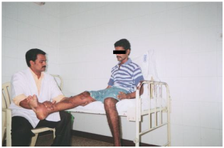
Fig.1 Subject performing post isometric relaxation
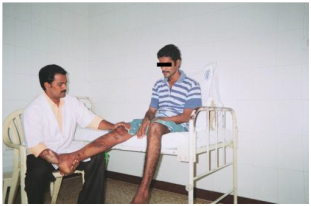
Fig.2 Subject performing reciprocal inhibition
Wax bath: The most widely used method of application for the extremity joints is “dip and wrap” method. The nature of wax treatment is explained and area to be treated is checked for contraindications the temperature of the wax also checked the treatment part is washed and thoroughly dried to prevent water being introduced in the wax bath. A patient is positioned to be able to wrap the part in wax in a convenient and comfortable way. The bandages of suitable size and mesh can be soaked in hot wax and then wrapped around the joint; the additional wax can then be brushed over the bandages. There are six to twelve wrappings used alternatively 12, 13 .
Static isometric exercise for the knee: Three methods which may be used to obtain this contraction. The contraction is taught on the unaffected leg and is seen and felt by the patient who then attempts a similar contraction on the other side. In lying one hand gives compression on the sole of the foot and resists plantar flexion strongly while the other hand placed under the knee joint. The therapist put one hand on the muscles and the other under the patients heel the patient is then asked to feel the pressure and attempt to relive it by lifting the leg. No movement takes place but the muscles are thrown into a state of strong contraction. Active Assisted Knee Mobilization: Active assisted knee mobilization performed either the assistance given by the therapist or by the patient himself. Position the patient in supine lying, instructs the patient to initiate the motion by lifting up the involved knee with the normal foot. Another method is to position the patient in high sitting, asks the patient to bend his knee maximum the effort of the patient can be assisted by the therapist or by the patients opposite leg.
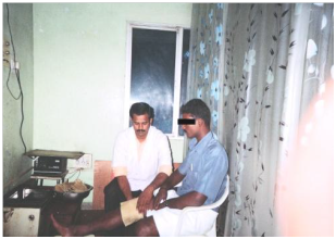
Fig.3 Subject receiving wax bath treatment
RESULT
Independent t test is used to comparing Post Test Vas Values Of Group A and Group B. On day 4 the post test VAS value of group A is 4.6 and group B is 5.8. the calculated t value is (10.18) is greater than the table value (t=2.048) at 5% level of significance for two-tailed test, showing that there is a significant difference between two groups, reject the null hypothesis. On day 7 the mean post test VAS value of group A is 2.2 and group B is 4.4. the calculated t value is (11.66) is greater than the table value (t=2.048) at 5% level of significance for twotailed test, showing that there is a significant difference between the two groups, reject the null hypothesis 14, 15 .
Dependent t test is used to comparing initial, day 4 and day7 vas values of Group A. The mean pre test VAS value is 6.8 and post test values are 2.2 and 4.6. Calculated t values are (20.9) & (35.88) is greater than the table value (t=2.145) at 5% level of significance for twotailed test, showing that there is a significant difference between the values. Dependent t test is used to comparing initial, day 4 and day 7 vas values within the Group B. The mean pre test VAS value is 6.9 and post test values are 1.1 and 2.5. Calculated t values are (16.5) & (19.5) is greater than the table value (t =2.145) at 5% level of significance for two-tailed test, showing that there is a significant difference between the values.
Independent t test is used to comparing post test active knee flexion values of Group-A and Group-B. On day 4 post test mean active knee flexion of group-A is 78 and group-B is 63.3. Calculated t value (4.831) is greater than the table value (t=2.048) at 5% level of significance for two-tailed test, showing that there is asignificant difference between two groups reject the null hypothesis. On day 7 post test mean active knee flexion of group-A is 103 and group-B is 78. Calculated t value (8.515) is greater than the table value (t=2.048) at 5% level of significance for two-tailed test showing that there is a significant difference between two groups reject the null hypothesis. Dependent t test is used compare initial, day 4 and day 7 values of Group-A. The mean pre test active knee flexion value is 56.3 and post test values are 21.7 and 46.7. Calculated t values are (16.15) & (27.2) is greater than the table value (t=2.145) at 5% level of significance for two-tailed test, showing that there is a significant difference between the values.
Dependent t test is used to comparing initial, day 4 and day 7 values of Group-B. The mean pre test active knee flexion value is 50.3 and post test values are 13.3 and 27.7.Calculated t values are (11.4) & (18.045) is greater than the table value (t=2.145) at 5% level of significance for two-tailed test, showing that there is a significant difference between the values. Independent t test is comparing post test quadriceps strength values of Group-A and Group-B. On day 4 post test mean of quadriceps strength of group-A is 5.8 and group-B is 4.9. Calculated t value (16.11) is greater than the table value (t=2.048) at 5% leve of significance for two-tailed test, showing that there is a significant difference between two groups reject the null hypothesis. On day 7 post test mean of quadriceps strength of group-A is 7.7 and group-B is 6.2. Calculated t value (6.0) is greater than the table value (t=2.048) at 5% level of significance for twotailed test, showing that there is a significant difference between two groups.
Dependent t test is used to compare initial, day 4 and day 7 values of Group-A. The mean pre test quadriceps strength value is 3.9 and post test values are 1.9 and 3.8. Calculated t values are (12.35) & (20.6) is greater than the table value (t=2.145) at 5% level of significance for two-tailed test, showing that there is a significant difference between the values. Dependent t test is used to compare initial, day 4 and day 7 values of Group-B. The mean pre test quadriceps strength value is 3.7 and post test values are 1.2 and 2.5. Calculated t values are (14.3) & (19.5) greater than the table value (t=2.145) at 5% level of significance for twotailed test, showing that there is a significant difference between the values.
Independent t test used to compare post test hamstring strength values of Group-A and Group-B. On day 4 post test mean of hamstring strength of group-A is 6.3 and group-B is 5.2.Calculated t value (5.714) is greater than the table value (t=2.048) at 5% level of significance for two-tailed test, showing that there is a significant difference between the two groups reject the null hypothesis.
On day 7 post test mean of hamstring strength of groupA is 8 and group-B is 6.4, Calculated t value (t=7.222) is greater than the table value (t=2.048) at 5% level of significance for twotailed test, showing that there is a significant difference between two groups. Dependent t test is used to comparing initial, day 4 and day 7 values of Group-A.the mean pre test hamstring strength value is 4.3 and post test mean values are 2 and 3.7. calculated t values are (26.0) & (24.45) greater than the table value (t=2.145) at 5% level of significance for two-tailed test, showing that there is a significant difference between the values.
Dependent t test is used to compare initial, day 4 and day 7 values of Group-B. The mean pre test hamstring strength value is 4 and post test values are 1.2 and 2.4. Calculated t values are (11.7) & (18.72) greater than the table value (t=2.145) at 5% level of significance for twotailed test, showing that there is a significance between the values.
DISCUSSION
Analysis of mean change in pain, active knee flexion and mean strength changes of both quadriceps and hamstrings revealed that there is significant difference between group-A who received muscle energy technique, wax bath, static quadriceps exercises and active assisted mobilization exercises, when compared with the group-B who received wax bath, static quadriceps exercises and active assisted mobilization exercises. Results obtained after analysis shows that there is decrease in pain score and increase active knee flexion, increase muscle strength respectively, which is statistically significant in post operative fracture stiffness patients who received muscle energy techniques when compared with the control group at the end of day 4 and day 7.
This permits rejection of null hypothesis. Analysis of results between pretest and post test values of experimental group shows that there is improvement in pain score, active knee flexion, and quadriceps, hamstrings strength following muscle energy techniques at the end of day 4 and day 7. Analysis of results between pretest and post test values of control group shows that there is significant improvement in pain score, active knee flexion and muscle strength following standard treatment at the end of day4 and day7. Hence the post analysis of results shows the superiority of muscle energy techniques along with standard treatment to reduce pain and increase range of motion and muscle strength. The better result in experimental group could be due to the muscle energy techniques decrease muscular spasm, reduce muscular shortening, prevents inter fiber adhesions influences the greater reduction of pain, increase range of motion, isometric exercise nature of this technique at various angle of restriction increase strength throughout the range of motion 16, 17.
Reason for decrease of pain and increase of range of motion, strength of muscle by muscle energy techniques: The most important causative factor for limiting joint range of motion after an injury is muscular shortening due to muscle spasm (Bourdilon, 1982).The restriction which takes place as a result of tight, shortened muscles usually accompanied by some degree of lengthening and weakness of antagonist, muscle energy technique targets these muscle tissues to promote relaxation, increase circulation, affecting Gamma motor neuron system in order to reduce pain and spasm 18.
In muscle energy technique a combination of both Post isometric relaxation and Reciprocal inhibition can effectively be employed to lengthen the shortened tissues and to strengthen the weak overlong muscles. When a muscle is isometrically contracted its antagonist will be inhibited and relaxed, similarly the agonist or shortened muscle also inhibited to achieve a degree of ease and additional movement of the shortened tissue 19 .
CONCLUSION
The study concluded that the standard treatment and standard treatment along with muscle energy technique both have an effect to reduce pain, increase range of motion, and strength of muscle after post immobilization fracture stiffness. The study also concluded that the Muscle energy technique was producing greater improvement in gaining range of motion and strength of muscles than other standard treatments.
REFERENCES
- J.Maheshwari, (1997). Essential Orthopaedics, 2nd revised edition, interprint, New delhi.
- Leon chaitow, et al. (2006). Muscle energy techniques Churchil Livingston, Singapore publishers (Pvt) Ltd.
- Carolyn kishner, Lymn Allen golby, Therapeutic exercise, 3rd edition, Jaypee brothers New delhi. (1996)
- M.Natarajan, N.Mayilvahanan, (1994). Orthopaedics and Traumatology, 4th edition, published by M.N. Orthopaedic hospital.
- Jayant joshi and Prakash kotwal (1999). Essential of orthopaedics and applied physiotherapy B.I.Publications New delhi.
- Susan.B.O’Sullivan (2001). Physical rehabilitation assessment and treatment, Jaypee brothers 4th edition, New delhi.
- David J. Magee (1997). Orthopaedic physical therapy assessment, 4th edition C.V.Mosby, Company, philadelphia.
- M.Dena gardener, (1985). Principles of exercise therapy, 4th edition, CBS publishers & distributors, New delhi.
- Leon chaitow (1993). Integrated neuromuscular inhibition techniques in the treatment of pain and trigger points.
- Cynthia C.Norkin, (2001). Joint structure and function, 3rd edition, Jaypee brothers New delhi.
- Cynthia C.Norkin, (1998). Measurement of joint range of motion, Jaypee brothers New delhi.
- John low, Ann Reed (1990). Electrotherapy explained, 3rd edition, ButterworthHeinmann, Oxford.
- Barbara J. Behrens, Physical agents, (1959). F. A. Davis company Philadelphia; 45.
- Kothari C.R., (2004). Research methodology, 2nd edition, new age international (pvt) ltd. New delhi.
- P.S.Sundar Rao and J.Richard, (2004). An introduction to biostatistics, 3rd edition, Prentice hall of India, New delhi.
- Lewit.K, Simons D.G., (1984). Myofascial pain; relief by post isometric relaxation, Archieves physical medicine and rehabilitation.
- Lewit.K, (1985). Muscular and articular factors in movement restriction, manual medicine.
- Liebenson C (1989). Active muscular relaxation techniques, journal of manipulative and physiological therapeutics.
- Lewit. K. (1986). Post isometric relaxation in combination with other methods of muscular facilitation and inhibition in manual medicine.
Citation:
Moorthy A, Jibi Paul, G Muthuraj (2019). Effects of muscle energy techniques on knee joint mobilization in an early stage following fracture shaft of femur and upper tibia , ijmaes, 5(1), 518-525.
Leave a Reply