

| Jibi Paul*1, Dainy Varghese2, Anitha Susan Cheriyan3 Corresponding Author: *1Professor and HOD, Department of Physiotherapy, Travancore College of Allied Health Sciences, Thattamala, Kollam, Kerala, India, Mail id: physiojibi@gmail.com Co-Authors: 2Professor and Principal, Travancore College of Allied Health Sciences, Thattamala, Kollam, Kerala, India 3Associate Professor, Department of Physiotherapy, Travancore College of Allied Health Sciences, Thattamala, Kollam, Kerala, India |
Abstract
| Background and Objective: Neck pain is a common musculoskeletal problem among dental professionals and may interfere with occupational activities, resulting in reduced functional capacity and increased disability. Prolonged and sustained postural demands during dental procedures may contribute to the development of mechanical neck pain. This study aimed to evaluate the effectiveness of an early isometric strengthening exercise program in reducing pain and improving functional ability among dental professionals with mechanical neck pain. Methods: A total of 30 dental professionals with mechanical neck pain participated in this comparative study. Participants were equally allocated into an experimental group and a control group, with 15 participants in each group. The experimental group received an isometric strengthening exercise protocol, whereas the control group received conventional physiotherapy incorporating interferential therapy with free neck exercises. The interventions were administered according to the study protocol, and changes in pain and functional ability were assessed before and after the intervention. Results: Both groups demonstrated improvement following the intervention. However, the experimental group receiving isometric strengthening exercises showed a statistically significant reduction in neck pain compared with the control intervention. The findings support the effectiveness of isometric strengthening exercises in reducing pain and improving functional outcomes among dental professionals with mechanical neck pain. Conclusion: An early isometric strengthening exercise program appears to be an effective intervention for reducing mechanical neck pain and improving functional ability among dental professionals. Incorporating targeted isometric strengthening exercises into physiotherapy management may help address postural musculoskeletal problems associated with dental practice. Keywords: Dental Professionals; Mechanical Neck Pain; Postural Neck Pain; Isometric Strengthening Exercises; Interferential Therapy; Physiotherapy |
| Received on 24th July 2026; Revised on 22nd August 2026; Accepted on 25th August 2026 DOI:10.36678/IJMAES.2026.V12I03.03 |
INTRODUCTION
Cervical spondyloarthrosis is a chronic degenerative condition affecting the cervical vertebrae, intervertebral discs, and associated structures of the neck. It is commonly characterized by intermittent or persistent neck pain, particularly among middle-aged and older adults. The intervertebral discs function as shock-absorbing structures between adjacent vertebrae. With advancing age, degenerative changes occur within the disc, including dehydration and loss of elasticity of the nucleus pulposus, accompanied by structural changes in the surrounding vertebral tissues.
The vertebral column has a complex anatomical and biomechanical organization that facilitates stability while allowing movement. The cervical spine is the most mobile region of the vertebral column and is therefore particularly susceptible to mechanical stress. Its extensive range of motion, combined with relatively limited structural support, may predispose the cervical region to mechanical and degenerative changes. These alterations can contribute to neck pain, stiffness, and functional discomfort, which are commonly associated with cervical spondylo-arthrosis¹.
Musculoskeletal discomfort associated with prolonged or sustained postures is frequently observed in individuals performing static occupational activities. Repeated exposure to awkward or sustained postures may result in cumulative mechanical stress involving the cervical spine and shoulder region, potentially leading to persistent pain, functional limitations, and disability. Several studies have identified neck, shoulder, and back pain as important occupational health concerns among dental professionals. The transition from predominantly standing-based dental practice to prolonged seated procedures has further increased the prevalence of musculoskeletal symptoms, particularly in the neck and shoulder regions².
Degenerative changes in the cervical spine may also involve the facet joints located posteriorly on either side of the vertebral canal. Progressive degeneration can result in the formation of osteophytes, or bony outgrowths, which may narrow the intervertebral foramina and irritate or compress adjacent nerve roots. Such changes can contribute to localized neck pain, radiating symptoms, and neurological irritation³.
The trapezius muscle is an important muscle connecting the upper limb to the axial skeleton and is particularly relevant to postural control of the cervical and shoulder regions. It is a broad, triangular muscle extending across the posterior neck and upper back. The muscle originates from the external occipital protuberance, the medial portion of the superior nuchal line, the ligamentum nuchae, the spinous process of the seventh cervical vertebra, and the spinous processes of the thoracic vertebrae, with attachment to the corresponding supraspinous ligament. The trapezius contributes to movements and stabilization of the scapula and plays an important role in maintaining cervical and shoulder posture4.
A spinal vertebra has very complex anatomical and biomechanical arrangement. Cervical spine is the most mobile part of the spine. This mobility of the spine is dependent on the anatomical integration. The cervical spine has got more mobility and poor anatomical support and hence it may lead to pathological changes, which gives rise to pathological changes, which gives rise to pain and discomfort in the neck which in clinical terms is called Cervical Spondylo-arthrosis5.
Occasional pains from irregular stances and postures are to be expected while they are performing static work. However, when the pain becomes regular occurrence, cumulative damage to the spine (cervical region), shoulder, could arise leading to debilitating injuries or also disability. Several studies have indicated that neck, shoulder and back pain are major problems among dentists. Eventually, when dentistry changed from standing job to sit down task, musculoskeletal pain in neck and shoulder region became more prevalent. This may explain the eventual focus on this areas6-9.
These degenerative changes can also affect the facet joints that lie behind the either side of the vertebral canal. There may be extra bone growth on the vertebrae called as the osteophytes, which may press the nerve roots causing pain and irritation of the nerves 10-13.
Objectives:
- To evaluate the effectiveness of isometric neck-strengthening exercises in reducing pain among dentists with mechanical neck pain.
- To determine the effect of conventional physiotherapy with free neck exercises on pain and functional ability.
- To compare the effectiveness of isometric neck-strengthening exercises with conventional physiotherapy with free neck exercises in improving pain and functional outcomes among dentists.
Aims and Objectives: To find out the effectiveness of isometric neck strengthening exercise and conventional physiotherapy with IFT on reducing pain and improve functional abilities among dentists. And to compare the results obtained from the data collected from both the groups under the study.
METHODOLOGY
The study population consisted of male and female dentists aged 27–40 years working at Oxford Dental College, Bangalore. A total of 30 dentists who met the eligibility criteria were selected using a simple random sampling method and allocated equally into two groups, with 15 participants in each group. Group A served as the control group and received interferential therapy, whereas Group B served as the experimental group and received isometric neck-strengthening exercises.
Selection Criteria and Intervention: The inclusion criteria comprised dentists aged 27–40 years who reported neck pain for a duration of three months or more and had mild to severe disability based on the Neck Disability Index (NDI).
Participants in Group A received free neck exercises for 10 minutes per session, whereas participants in Group B performed isometric neck-strengthening exercises for 10 minutes per session. Both interventions were administered once daily, five days per week, for a period of four weeks.
Outcome Assessment: A pre-treatment assessment was conducted for both groups using the Visual Analogue Scale (VAS) to assess pain intensity and the Neck Disability Index (NDI) to evaluate the level of neck-related disability. The respective interventions were administered once daily, five days per week, over four consecutive weeks. Following completion of the intervention period, a post-treatment assessment was performed using the same outcome measures, namely the VAS and NDI, to determine changes in pain intensity and disability.
Data Collection: An assessment chart containing relevant information for all study participants was used for data collection. The chart facilitated the systematic recording and categorization of the information obtained from each participant. The collected data were subsequently organized and entered into a master chart for statistical analysis and interpretation.
RESULTS
The result analysis must be organized in such a way that all the questions can be answered. An interpretation was carried out in the later stage to make sure that the result can be implicated in a broader context.
Measurement tools for the study were Visual analogue scale, Neck disability index and Orthopaedic assessment chart. All the subjects both experimental and the control group were used the same orthopaedic assessment chart in order to maintain the validity and reliability of the research work.
| Group | Parameter | Time interval | Mean± SD | P-Value |
| Conventional Group | VAS | Pre | 7.63± 0.74 | <0.001 |
| Post | 6.13±0.93 | |||
| NDI | Pre | 34.04±1.88 | <0.001 | |
| Post | 37.75±1.46 | |||
| Experimental Group | VAS | Pre | 8.03±0.64 | <0.001 |
| Post | 3.67±0.72 | |||
| NDI | Pre | 35.6±1.35 | <0.001 | |
| Post | 42.75±1.62 |
Table 1: Comparison of various parameters between control & experimental groups
The main aim here is for interpretation of data collected from 30 patients who participated in the study. The data collected is tabulated carefully and master chart is made and the results are analyzed and interpreted using statical tools.
The results of this study are analyzed in inter and intra group by different statical tools. The comparative analysis of the data of level of pain and the lower extremity functional scale of the experimental and control group are compared between the pre and post treatment phases.
A statistically significant difference was observed between the control and experimental groups with respect to mean VAS scores in the post-treatment phase (P < 0.001). The control group demonstrated a higher mean VAS score than the experimental group, and this between-group difference was statistically significant.
Similarly, a statistically significant difference was observed in mean NDI scores between the control and experimental groups in the post-treatment phase (P < 0.001). The experimental group showed a higher mean NDI score compared with the control group, with the difference between the groups reaching statistical significance.
Within the control group, a statistically significant difference was observed in mean VAS scores between the pre-treatment and post-treatment phases (P < 0.001). The mean VAS score decreased following treatment, and this reduction was statistically significant. In contrast, the mean NDI score in the control group increased from the pre-treatment to the post-treatment phase, and this increase was also statistically significant (P < 0.001).
| Parameter | Group | Mean± SD | P-Value |
| VAS – Pre | Control | 7.63± 0.74 | 1.000 |
| Experimental | 8.03±0.64 | ||
| VAS – Post | Control | 6.13±0.93 | <0.001 |
| Experimental | 3.67±0.72 | ||
| NDI – Pre | Control | 34.04±1.88 | 0.233 |
| Experimental | 35.6±1.35 | ||
| NDI – Post | Control | 37.75±1.46 | <0.001 |
| Experimental | 42.75±1.62 |
Table 2 : Comparison of various parameters within each group between pre and post treatment phase
In experimental group, we notice that there is a significant difference in mean VAS between Pre and Post treatment phase (P<0.001). We notice that there is a decrease in mean VAS from pre-treatment phase to post treatment phase and the mean decrease is found to be statistically significant. Mean NDI in experimental group is found to have increased from pre treatment phase to post treatment phase and the increase is found to be statistically significant (P<0.001)
In control group, we notice that there is a significant difference in mean VAS between Pre and Post treatment phase (P<0.001). We notice that there is a decrease in mean VAS from pre-treatment phase to post treatment phase and the mean decrease is found to be statistically significant.
Mean NDI in control group is found to have increased from pre-treatment phase to post treatment phase and the increase is found to be statistically significant (P<0.001).
In experimental group, we notice that there is a significant difference in mean VAS between Pre and Post treatment phase (P<0.001). We notice that there is a decrease in mean VAS from pre-treatment phase to post treatment phase and the mean decrease is found to be statistically significant.
Mean NDI in experimental group is found to have increased from pre-treatment phase to post treatment phase and the increase is found to be statistically significant (P<0.001)
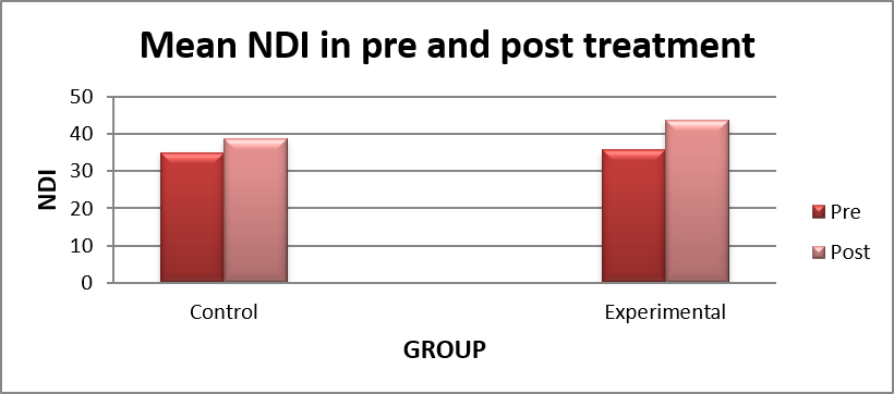
Graph1: Mean difference in NDI on both Groups
DISCUSSION
The present study was conducted to evaluate the effectiveness of an early isometric strengthening intervention in individuals with mechanical neck pain, in comparison with a conventional treatment protocol.
A total of 30 subjects participated in the study and were randomly allocated into two groups: a control group and an experimental group. The control group received interferential therapy (IFT) for a period of four weeks, whereas the experimental group underwent an early isometric neck-strengthening intervention during the same treatment period.
Pre- and post-treatment assessments were performed using a general physical examination and detailed history taking. In addition, standardized outcome measures, namely the Visual Analogue Scale (VAS) and Neck Disability Index (NDI), were used to evaluate pain intensity and functional disability, respectively. These outcome measures provided the primary basis for statistical comparison between the control and experimental groups.
The statistical analysis included the mean, standard deviation, t-value, and p-value for both VAS and NDI scores. The assessment was performed during the pre-treatment and post-treatment phases for both groups, allowing changes following the respective interventions to be evaluated.
The findings demonstrated that there was no statistically significant difference between the control and experimental groups during the pre-treatment phase, indicating that the groups were comparable with respect to pain intensity and functional disability at baseline. However, a statistically significant difference was observed between the groups in the post-treatment phase, with the experimental group demonstrating greater improvement in the outcome measures.
The statistical findings therefore indicate that the early isometric strengthening intervention was effective in reducing pain and improving functional ability in individuals with mechanical neck pain. As the observed post-treatment differences were statistically significant at the predetermined level of significance (p < 0.05), the null hypothesis was rejected and the alternative hypothesis was accepted.
Overall, the results suggest that early isometric strengthening may be an effective therapeutic intervention for reducing pain and improving functional outcomes in individuals with mechanical neck pain.
The present study was undertaken to evaluate the effectiveness of early isometric strengthening exercises in individuals with mechanical neck pain and to compare their effects with those of a conventional treatment protocol using interferential therapy (IFT). A total of 30 subjects participated in the study and were randomly allocated into a control group and an experimental group. Pain intensity and functional disability were assessed before and after the intervention using the Visual Analogue Scale (VAS) and Neck Disability Index (NDI), respectively.
The findings of the present study demonstrated that there was no statistically significant difference between the control and experimental groups during the pre-treatment assessment. This indicates that the two groups were comparable at baseline with respect to pain intensity and functional disability. The absence of a significant baseline difference is important because it suggests that the subsequent differences observed following treatment were more likely to be associated with the respective interventions rather than substantial pre-existing differences between the groups.
Following four weeks of intervention, a statistically significant difference was observed between the groups in both VAS and NDI scores. The experimental group, which received early isometric strengthening exercises, demonstrated greater improvement compared with the control group. The reduction in VAS scores indicates a decrease in perceived pain, while improvement in NDI scores indicates a reduction in neck-related functional disability. These findings suggest that incorporating early isometric strengthening into the management of mechanical neck pain may provide beneficial effects on both symptoms and functional performance.
The improvement in pain observed in the experimental group may be explained by the effects of isometric muscle activation on neuromuscular function. Isometric exercises involve muscle contraction without substantial joint movement and can be performed while placing relatively controlled mechanical demands on the cervical region. Progressive activation of the cervical musculature may improve muscle recruitment, endurance, and dynamic support of the cervical spine. Improved muscular control may subsequently reduce excessive mechanical stress on cervical structures during daily activities and sustained postures, thereby contributing to a reduction in pain.
Another possible explanation for the reduction in pain is the influence of therapeutic exercise on pain modulation. Muscle contraction can stimulate sensory afferent input and may contribute to endogenous pain-inhibitory mechanisms. Repeated controlled contractions may therefore influence pain perception and tolerance in individuals with mechanical neck pain. However, the exact mechanisms responsible for the observed clinical improvement cannot be established solely from the present study and may involve several interacting physiological and biomechanical factors.
The improvement in NDI scores in the experimental group is particularly relevant because neck pain is frequently associated with limitations in activities of daily living. Pain may interfere with activities such as prolonged sitting, reading, working at a computer, driving, lifting, and maintaining sustained neck postures. By improving cervical muscle strength and endurance, isometric exercises may enhance the individual’s ability to tolerate these activities. Thus, the reduction in disability observed following the strengthening intervention may reflect not only decreased pain but also improved functional capacity.
The findings also indicate that early strengthening may have advantages over a treatment approach directed primarily toward symptomatic relief. IFT is commonly used as a physical therapy modality for pain management and may provide short-term symptomatic benefits. Its proposed effects are associated primarily with modulation of pain perception through electrical stimulation. However, IFT does not directly provide the same progressive muscular loading and neuromuscular training achieved through active strengthening exercises. Therefore, the greater improvement observed in the experimental group may be related to the active nature of the intervention and its potential effects on muscle performance and cervical stability.
An important aspect of the present study is the use of early isometric strengthening rather than delaying strengthening until symptoms have completely resolved. Traditionally, exercise progression may be approached cautiously in individuals experiencing pain. However, appropriately dosed isometric exercise can provide a relatively controlled method of introducing muscle activation while minimizing excessive cervical movement. Early activation may help prevent prolonged muscular deconditioning and may facilitate a gradual return to normal functional activities.
The results of the present study also have potential clinical implications. Mechanical neck pain is common among individuals exposed to prolonged static postures and repetitive occupational activities. Management strategies that combine pain reduction with restoration of muscular function may therefore be particularly useful. The present findings suggest that early isometric strengthening could be considered as part of a rehabilitation programme rather than relying exclusively on passive modalities for symptom management.
The significant difference observed in the post-treatment VAS scores indicates that the experimental intervention was associated with a greater reduction in pain compared with the control intervention. From a clinical perspective, reduction in pain may allow patients to participate more actively in rehabilitation and daily activities. Similarly, the significant improvement in NDI scores suggests that the intervention may have contributed to better functional performance. The simultaneous improvement in both pain and disability strengthens the interpretation that the intervention may have produced benefits extending beyond pain relief alone.
The present findings are also consistent with the general principle of active rehabilitation, in which restoration of strength, endurance, mobility, and neuromuscular control is considered important for the management of musculoskeletal disorders. Exercise-based interventions may address some of the underlying physical impairments associated with mechanical neck pain, whereas passive modalities may primarily target symptoms. Therefore, the greater post-treatment improvement in the experimental group may support the incorporation of active strengthening strategies into physiotherapy management.
The absence of a significant difference between the groups during the pre-treatment assessment further supports the validity of the post-treatment comparison. Since both groups began with broadly comparable levels of pain and disability, the statistically significant difference following treatment provides evidence that the experimental intervention was associated with greater improvement. Nevertheless, statistical significance should be interpreted together with the magnitude of change and clinical relevance of the observed improvement.
The findings of the present study support the alternative hypothesis that early isometric strengthening is effective in reducing pain and improving functional ability in individuals with mechanical neck pain. Consequently, the null hypothesis was rejected at the predetermined significance level of p < 0.05. The results suggest that early strengthening may be a useful component of conservative physiotherapy management for mechanical neck pain 12.
The findings have practical implications for physiotherapy practice. Early introduction of controlled isometric strengthening exercises may provide an active treatment option for individuals with mechanical neck pain. Such exercises may be particularly useful when the clinical objective is not only to reduce pain but also to restore cervical muscle function and improve tolerance to daily activities. The intervention can potentially be incorporated alongside other appropriate physiotherapy approaches based on individual patient presentation13.
Several limitations should be considered when interpreting the findings of the present study. The sample size was relatively small, with only 30 participants, which may limit the generalizability of the findings to a larger population. The intervention period was limited to four weeks, and therefore the long-term effectiveness and maintenance of the observed improvements could not be determined. In addition, the study relied primarily on VAS and NDI as outcome measures. Although these are widely used clinical measures, additional objective measures such as cervical muscle strength, endurance, range of motion, and postural parameters could have provided further information regarding the mechanisms underlying the observed improvements14.
The study population was also restricted to a specific group of individuals, which may limit the applicability of the findings to patients with different demographic or occupational characteristics. Furthermore, factors such as adherence to the prescribed exercise programme, ergonomic habits, physical activity levels, and psychosocial influences may also affect the outcome of mechanical neck pain and should be considered when interpreting the results15,16.
Future studies should include larger sample sizes and longer follow-up periods to determine whether the benefits of early isometric strengthening are maintained over time. Randomized controlled trials with additional outcome measures, including objective assessments of cervical muscle strength, endurance, range of motion, and functional performance, would provide a more comprehensive understanding of the effects of the intervention. Future research may also compare early isometric strengthening with other active exercise approaches or with combined treatment protocols involving both therapeutic exercise and electrotherapy17,18.
Overall, the findings of the present study indicate that early isometric strengthening exercises produced greater improvements in pain and functional disability than the conventional IFT-based treatment protocol used in the control group. The statistically significant post-treatment differences in VAS and NDI suggest that early strengthening may be an effective component of conservative management for mechanical neck pain. The findings further emphasize the importance of active rehabilitation in addressing both symptoms and functional limitations associated with mechanical neck pain19.
CONCLUSION
Subjects with mechanical neck pain mainly suffer from pain and difficulty in their activities during course of the disease. This study was aimed at determining or finding out the effectiveness of the early isometric neck strengthening in dental personals with mechanical neck pain. In this study, 30 subjects with mechanical neck pain actively participated, 15 of whom underwent early isometric strengthening intervention in the experimental group.
On the basis of the data obtained it clearly showed that there was clear reduction in the pain and disability level in the experimental group and control group, but when both the groups were compared experimental group showed better improvement than control group. Therefore, going by the overall result of the study it can be concluded that, early isometric strengthening is indeed effective in reducing pain and improving functional ability in dental professionals with mechanical neck pain.
REFERENCES
- Mc Gill.S, Hughson, R., Parks, K. (2000). Lumbar erector spinae oxygenation during prolonged construction: implications for prolonged work. Ergonomics,43,486-493
- Ylippa v.,szuster, F., Spencer, J.,preber,H., benko, S., Arnetz, B.(2000). Health, mental well-being and musculoskeletal disorders:a comparison between Swedish and Australia dental hygienists. Journal of dental hygiene, winter 76, 47-59
- Lehto, T., helenius,H. And alaranta,H.(1991). Musculoskeleton symptoms of dentists assessed by a multidisciplinary approach. community dentistry and oral epidemiology, 19,38-44
- Chase it all chase 1 robertson VJ, southwell B, hutson J, Gibb S “pilot study usingtranscutaneous electrical stimulation (interferential current) to treat chronic resistant constipation and soiling in children Gastroentral Hepatic. Jul.2005;20(7):1054-1061
- Johnson MI, JG. A single-blind placebo-controlled investigation into the analgesic effects of IF currents on experimentally induced ischaemic pain in healthy subjects cli. Physio. Funct. Imaging.May 2002;22(3):187-196.
- Noble JG Henderson G.cramp AF , Walsh DM , Lowe AS. The effect of IFT upon cutaneous blood flow in humans. Clin. physio. Jan 2000;20(1):2-7
- Mirkin G (2006) “isometric exercise background” second journal rehab. Medicine 11(1);33-34 PMID
- Diakow.P, Cassidy.,D.(1984).” Back pain in dentist”.J Manipulative and Physiological Therapeutics, 7,85-88.
- Finsen.L, Christensen.H , Bakke.M (1998) “Musculoskeletal disorders among dentists and variation in dental work.”Applied Ergonomics,29,119-125.
- Gray.H (2000) “Anatomy of the human body”.Philadelphia;Lea and Febigei,1918;Bartleby.com (2004)
- Murtomaa.H (1982) “Work-related complaints of dentists assistants”.International Archives of Occupation and Environmental Health,50,231-236.
- Runderantz.B,Johnson.B(1990) “Cervical pain and discomfort among dentists”.Epidemiological,clinical and therapeutic aspects,Swedish dental journal,14,71-80.
- Ratzon.N,Yaros.T,Mizlik(2000) “Musculoskeletal symptoms among dentists in relation to work posture.” Work,15,153-158.
- Valchi.B, Valichi.K(2003) “Mechanisms leading to musculoskeletal disorders in dentistry.” J American Dent.Assoc,134,1344-1350.
- Neubaver.J, Zablten-Hinguranage A, Schilten Woft M, Boucher M- multimobal therapy in patients with chronic cervical and lumbar pain, results of a comparative prospective study 2005 Sep 8 (2) 10-15.
- Crowford J R, Khanb R J, Varly G W – early management outcome following soft tissue injuries of the neck A randomized controlled trail Jour of Spine Injuries 2004 Sep:35(9): 891-5.
- Nadler.SF (2004). “Forceanalysis of individual muscle of the shoulder joint during asometric exercise”. J Biomechanics.6,385-393.
- Sarig Bahat H. (2003). “Evidence of exercise thearapy in mechanical neck disorders.” Dept of Physiotherapy Isreil,2003 Feb;8(1);10-20.
- Y.Chui, Lam TH, Hedly (2003) “A randomized controlled trial on exercises for patients with chronic neck pain.”Aug(1493)605-627, Phy Med Rehab Cli. M Amc.
| Citation: Jibi Paul, Dainy Varghese, Anitha Susan Cheriyan (2026). Comparative effectiveness of Isometric Strengthening Exercises and Conventional Physiotherapy in the management of Mechanical Neck Pain Among Dental Professionals, ijmaes, 12(3), 2897-2907. |
Leave a Reply