

Nabilah Ahmad1*, Siti Aishah Abdullah Suhaimi2, Najiah Anuar2, Dinesh Madhavan Nair2, SitiNurBaait Sokran1
Authors:
1Physiotherapy Department, School of Health Sciences, KPJ Healthcare University College, 71800 Nilai, Negeri Sembilan, Malaysia.
2Medical Imaging Department, School of Health Sciences, KPJ Healthcare University College, 71800 Nilai, Negeri Sembilan, Malaysia.
Corresponding Author:
1*Physiotherapy Department, School of Health Sciences, KPJ Healthcare University College, 71800 Nilai, Negeri Sembilan, Malaysia. Mail id: ucn.nabilah@kpjuc.edu.my
ABSTRACT
| Background and objectives: Dynamic warm-ups prepare the body for activity by helping to increase blood flow and muscle temperature. By calculating the muscle elongation, muscle thickness and pennation angle, it will show the effectiveness of the dynamic elongation task. Ultrasound imaging involves the use of a transducer (probe) and ultrasound gel placed directly on the skin. Ultrasound images of the musculoskeletal system provide the pictures of muscles, tendons, ligaments, joints, and soft tissues throughout the body. Therefore, this study aimed to determine the changes in the muscle tendon unit displacement among healthy male subjects in dynamic task of a gastrocnemius muscle. Methods: This experimental studywas participated by 32 healthy male subjects among KPJUC students. Musculoskeletal Ultrasound (MSK Ultrasound) performed to collect the databefore and after the dynamic task. The measurement was taken for pre and post dynamic elongation task. Paired sample t-test and paired samplecorrelation were used as a statistical analysis. Results: This study shows that there is a changes in muscle architecture after the dynamic elongation task. There is significant difference in pennation angle and muscle elongation between pre dynamic elongation task and post dynamic elongation task. For muscle thickness, there is no significant different between pre dynamic elongation task and post dynamic elongation task. Conclusion: There is a change in muscle tendon unit displacement for gastrocnemius muscle between pre dynamic elongation task and post dynamic elongation task and the obvious changes can be seen in pennation angle of the muscle. Dynamic elongation task seems to be an effective stretching for rehabilitation purposes because it can produce the changes in muscle architectures. |
Keywords: MSK Ultrasound, Pennation Angle, Muscle Thickness, Muscle Elongation, Dynamic Stretching
| Received on 20th February 2020, Revised on 26th February 2020, Accepted on 29th February 2020. DOI:10.36678/ijmaes.2020.v06i01.007 |
INTRODUCTION
Abnormal muscle tendon elongation occurs when the injury to the muscle happens. For management and prevention of the injuries there is an important components to understand of muscle tendon elongation. During any sort of movement, muscle tendon unit is the one which generates force production of a particular muscle 1.
The force production can be either active or passive force, which relies on length of the muscle. It is based on the length amount of sarcomeres will be recruited. There is no previous study examined the pattern of elongation and structural changes at the level of muscle tendon unit. It is believed that understanding such mechanism of muscle tendon unit explains the science behind the injury mechanism. The regular elongation to a muscle contribute to a defined movement of muscle tendon and joints 2, 3.
Ultrasonography is a valid tool which shows any changes in muscle tendon length properties. The drawback of the usage of ultrasonography tool is its unclear how the elongation mechanism occurs in dynamic elongation. Therefore, uncertain prevails on types of elongation task is required for rehabilitation outcome. Thus, there is a need to understand the elongation mechanism for dynamic task on a muscle. Muscle imaging was used to show that the ultrasonography could properly estimate muscle activity. They measured architectural parameters which included the pennation angle, fascicle lengths and the muscle thickness. Ultrasonography is used to understand biological and bioelectrical characteristics of muscle. An ultrasound is a proper non-invasive real time imaging for muscle structures. Collected data will answer properties of the muscle tendon unit elongation mechanism through displacement of the tendon. This study prescribes either of the elongation task for a variety of patients as well for normal subjects in order to improve social well-being 4, 5.
METHODOLOGY
This experimental study was conducted in KPJ Healthcare University College (KPJUC), Nilai. A total of 32 healthy individuals was recruited and subjected to undergo the dynamic elongation technique with enough rest periods. The normal healthy individuals for this study was identified among the students who are studying in KPJUC. The subject recruitment were based on the established inclusion criteria.
The measurement was taken for pre dynamic elongation task and post dynamic elongation task. Real time ultrasound imaging (Mylab Touch, Esaote, Italy) 15-MHz linear type probe with 38 mm wide field of view (FOV) were used to measure tendon displacement, muscle thickness, pennation angle and muscle elongation. Another tool is treadmill machine, which is used to do the warming up maneuver and the metronome for monitoring the number of beats while performing dynamic elongation.
Subjects were asked to walk in the treadmill for 5 minutes as a warming up. Then, the subjects made to perform dynamic elongation on their dominant legs only then they were stand with dominant leg and to raise the entire foot off the floor, which lead to hip flexion. Then the subjects were instructed to perform active movement of foot to a rhythm of 60 beats per minutes (60 BPM) with the help of metronome and each movement was performed for 1 second. The dynamic elongation was done for 30 second and will be repeated for 5 times. Elongation maneuver pre and post measurement of the subject’s muscle-tendon unit displacement, fascicle length and pennation angle were obtained.
The measurement starting on 30 mm below the fossa popliteal and about 20 mm medial of the line separating the medial and lateral gastrocnemius muscle. In this location the muscle fibers have a distinctly visible pennation angle and muscle structure seems to be well-define. Each subject instructed to stand upright with feet parallel, looking at the same point on the front wall. Prior to stretching, the middle of the monitor display was marked with a white string. A rectangular plastic foam frame (proximal frame) through which the ultrasound probe could pass was placed onto the right calf of each subject to obtain measurements from the same location, a quarter proximal to the distance between the popliteal crease and center of the lateral malleolus.
Myotendinous junction (MTJ) was defined as where the superficial and deep aponeuroses of medial gastrocnemius (MG) met. Another rectangular plastic foam frame (distal frame) was put on the right calf where the middle of the MTJ of the MG aligned with the midline of the ultrasound monitor, which was defined as the baseline of the MTJ. After the dynamic stretching, the probe was set in the same place and the image was taken. The MTJ then calculated by measuring the distance between the white reference line and the new MTJ position. The proximal displacement of the MTJ will show in ultrasound image. The pennation angle of the MG and fascicle length (Lf) were also assessed from the images, which were taken at the proximal frame. The pennation angle of MG was measured as the angle of insertion of the muscle fiber fascicles into deeper aponeurosis. Fascicle length (Lf) was defined as the length of the fascicular path between the insertions of the fascicle into the upper and deeper aponeuroses.
RESULTS
A total of 32 healthy young man participated in the study. The demographic data obtained include male subjects who are aged between 20 – 25 years old. The male subjects who does not have any lower limb injury such as ligament or muscle tear and who have normal Body Mass Index which in range 18.5-24.9𝑘𝑔/𝑚2. Subjects was categorized into two groups; right dominant leg and left dominant leg.
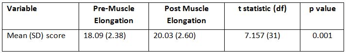
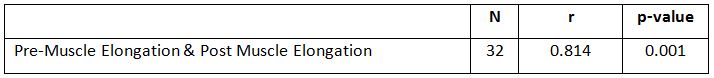
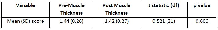
Majority of the healthy young man participated were right dominant leg (94%), and the remaining healthy young man were left dominant leg (6%). The p value for muscle elongation (p=0.00) which is<0.05, therefore reject the null hypothesis and there is a significant difference. There is a significant difference of mean score between Pre Muscle Elongation and Post Muscle Elongation after an intervention. The significant relationship of score between Pre Muscle Elongation and Post Muscle Elongation which is strong (0.814).The p value for muscle thickness (p = 0.606)>0.05, therefore not reject the null hypothesis and there is no significant difference.
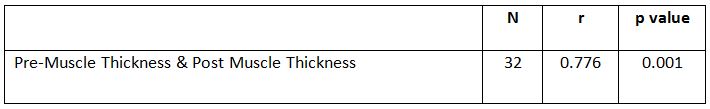
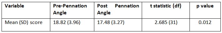
There is no significant difference of mean score between Pre Muscle Thickness and Post Muscle Thickness after an intervention. The significant relationship of score between Pre Muscle Thickness and Post Muscle Thickness, which is strong (0.776).The p value for pennation angle (p = 0.012)<0.05, therefore reject the null hypothesis and there is a significant difference. There is a significant difference of mean score between Pre Pennation Angle and Post Pennation Angle after an intervention.
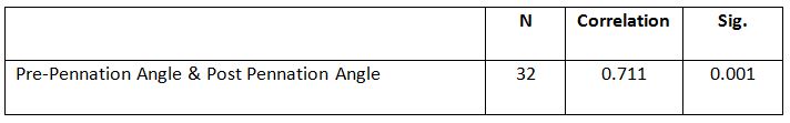
The significant relationship of score between Pre Pennation Angle and Post Pennation Angle, which is strong (0.711).
DISCUSSION
The age of subjects was fixed in the range of 20 to 25 years old because of the composition of Skeletal Muscle Mass might be stable during the age of 20 to 40 years old and at the age of 45 years old it begins to decrease significantly. Due to decreases in the amount and diameter of muscle fibers it caused the decrease in Skeletal Muscle Mass occurs with aging process as a physiological change. Dominant leg for the subjects also have to consider because of the scanning need to be done on the dominant leg. Leg dominance has been determined by which hand dominant is dominant. If the person is left-handed, the he must be left leg dominant6.
The definition of muscle power is the amount of work a muscle can produce per unit of time. High muscle power understood as the capacity to exert high levels of strength as quickly an explosively as possible. No statistical difference in maximal power between the dominant and non-dominant legs in healthy young adults, whether they are non-athletes or professional, single-leg-dominant athletes and the reason younger group of healthy man was chosen in my study is because muscular power development reaches its peak between 18 and 30 years of age, so theoretically I had the best chance to find asymmetries in this age range7.
The results shows the dynamic elongation task is an effective stretching since there is a different length of gastrocnemius muscle between pre and post, this results supported by the study of Knudson et al., 2006 which is when a muscle or muscle group is passively stretched using techniques like in static, dynamic, or proprioceptive neuro-muscular facilitation (PNF) stretching there might be some short-term changes in the muscle. The short-term or acute effects of stretching on muscle relate to the initial performance changes in the first few hours after stretching.
Therefore, the acute effects following stretching then depends on the biomechanical performance variables like a range of motion (ROM) have been shown to improve following stretching, while some of it appear to be unaffected such as stiffness and others are significantly reduced which means strength. The acute effect of the stretching on flexibility is clear. Stretching an acute increase in joint range of motion that tends to persist for 60 to 90 minutes. For rehabilitation purposes, passive stretching of the injured muscle helps elongate the maturing inter-muscular scar and prepares the muscle for strengthening. Dynamic training exercises can be added in a consecutive manner as each type of exercise is completed with painless to the patient8.
The muscle thickness slightly decreased after stretching was performed. A study from Simpson, Kim, Bourcet, Jones &Jakobi, et al. (2017) main findings were novel to human stretch training studies and included an increase in the thickness of gastrocnemius muscle, and increase in the fascicle lengths at both the MTJ and muscle belly with extent of the lengthening greater in the lateral gastrocnemius muscle compared with medial gastrocnemius muscle. The findings were contradict with the results from this study where the muscle thickness was slightly decreased.
The pennation angle was slightly decreased after the dynamic elongation task was performed. A review of literature of pennation angle and fascicle length of human skeletal muscles to predict the strength of an individual muscle using Real-Time Ultrasonography. found that The pennation angle defined as the pattern of arrangement of muscle fibers in relation to the axis of the force generation by the same muscle which is crucial component to determining muscle performance9.
The only study we found in the literature that investigating the effects of dynamic stretching exercises on muscle morphology demonstrated that dynamic stretching performed before exercise activities was not effective on fascicle length and pennation angle of the gastrocnemius muscle10.
In this study, the correlation between each parameters were not investigated. Therefore, it is recommended for future research to measure the correlation between each parameters. The age range of this study was limited from 20 years old to 25 years old, to overcome this limitation future study should wide the age gap.
Ethical Clearance: Received approval letter from the Research Ethics Committee, School of Health Sciences, KPJ Healthcare University College with reference number: KPJUC/RMC/ MPT/ EC/ 2018 /129 dated 19/03/2018.
Fund for the study: Research Management Center, Department of Physiotherapy, School of Health Sciences, KPJ Healthcare University College, Malasia.
Conflict of Interest: All authors have no conflict of interest to declare on conduct of this study.
CONCLUSION
The aim of this study is to determine the changes in the muscle tendon unit displacement among healthy male subjects in dynamic task of a gastrocnemius muscle. The data was collected on pre dynamic elongation task and post dynamic elongation task. The investigation of this study show that there is a changes in muscle tendon unit displacement for gastrocnemius muscle between pre dynamic elongation task and post dynamic elongation task and the obvious changes can be seen in pennation angle of the muscle. The results may be influence by subject BMI, height, weight and daily lifestyle. Moreover, for rehabilitation purposes, this dynamic elongation task seem to be an effective stretching because it can produce the changes in muscle architectures.
REFERENCES
- Hodges, P., Pengel, L., Herbert, R. and G andevia, S. (2003). Measurement of muscle contraction with ultrasound imaging. Muscle & Nerve, 27(6), 682-692.
- Vaisman, A., Guiloff, R., Rojas, J., Delgado, I., Figueroa, D., & Calvo, R. (2017). Lower limb symmetry: Comparison of muscular power between dominant and nondominant legs in healthy young adults associated with single-leg-dominant sports. Orthopaedic Journal of Sports Medicine, 5(12), 232-236.
- Knudson, Duane (2006). The biomechanics of stretching. Journal of Exercise Science and Physiotherapy, Vol. 2 : 3-12.
- Rekabizadeh M, Rezasoltani A, Lahouti B, Namavarian N.(2016). Pennation Angle and Fascicle Length of Human Skeletal Muscles to Predict the Strength of an Individual Muscle Using Real-Time Ultrasonography: A Review of Literature. J Clin Physio Res, 1(2): 42-48.
- Samukawa, M., Hattori, M., Sugama, N., & Takeda, N. (2011). The effects of dynamic stretching on plantar flexor muscle-tendon tissue properties. Manual Therapy, 16(6), 618-622.
- Miura, K., Yamamoto, M., Tamaki, H., &Zushi, K. (2010). Determinants of the Abilities to Jump Higher and Shorten the Contact Time in a Running 1-Legged Vertical Jump in Basketball. Journal of Strength and Conditioning Research, 24(1), 201-206.
- Wattimena, R., Vitriana, V., &Defi, I. (2017). Correlation between body mass index, gender, and skeletal muscle mass cut off point in Bandung. International Journal of Integrated Health Sciences,5(2), 47-51.
- Brukner, P., & Khan, K. Brukner& Khan’s (2002).Clinical sports medicine. Revised 2nd ed. McGraw-Hill, Australia.
- Zhou, G., Chan, P. and Zheng, Y. (2015). Automatic measurement of pennation angle and fascicle length of gastrocnemius muscles using real-time ultrasound imaging. Ultrasonics, 57, 72-83.
- Simpson, C., Kim, B., Bourcet, M., Jones, G., & Jakobi, J. (2017). Stretch training induces unequal adaptation in muscle fascicles and thickness in medial and lateral gastrocnemii. Scandinavian Journal of Medicine & Science in Sports, 27(12), 1597-1604.
| Citation: Nabilah Ahmad, Siti Aishah Abdullah Suhaimi, Najiah Anuar (2020). Effect of dynamic stretching on elongation of Gastrocnemius muscle, International Journal of Medical and Exercise Science, 6 (1): 713-719. |
Leave a Reply